Scofield Scientific Schematics and Cartoons

Everyone loves a great diagram, schematics or cartoon!
Something about these quirky bits of science and art has always fascinated me. This started off as an affinity to the figure images in text books, as they seemed to bring all the moving pieces together in a way that aided in the construction of a mental model of biological systems. As an independent investigator / educator, I have found these visual aids to be valuable tools for communicating results and teaching students and volunteers in the lab.
I think its fun to update them too, as results come in these visual representations become more and more detailed in our minds and in turn become more detailed on the page.
While Im certainly still learning and am by no means an expert on graphic design, I have collected some of the better drawings and cartoons that I have made and used to illustrate my work. I have selected images for this section that dont show up in the main project pages. Also, If the image was featured in a paper, I will link the original publication in the description below.

This is a recent attempt of mine to depict signaling across microglia, neurons and astroglia. The particular signal transduction cascade pictured is that of chronic pain and its cellular impacts on systems that overlap with relapse vulnerability. This one is not yet published. A variant of this template was used in a recent submission of a review article. I will post the link when the paper is published.
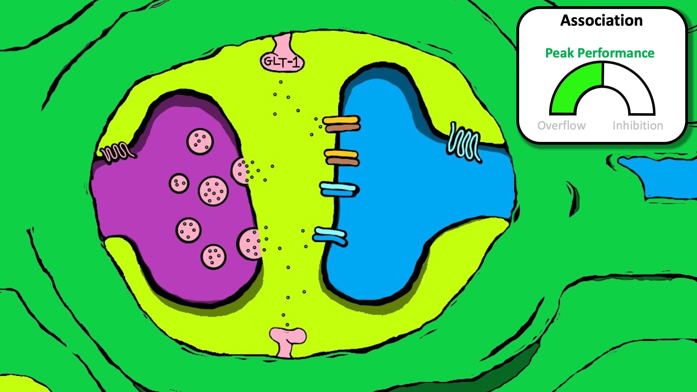
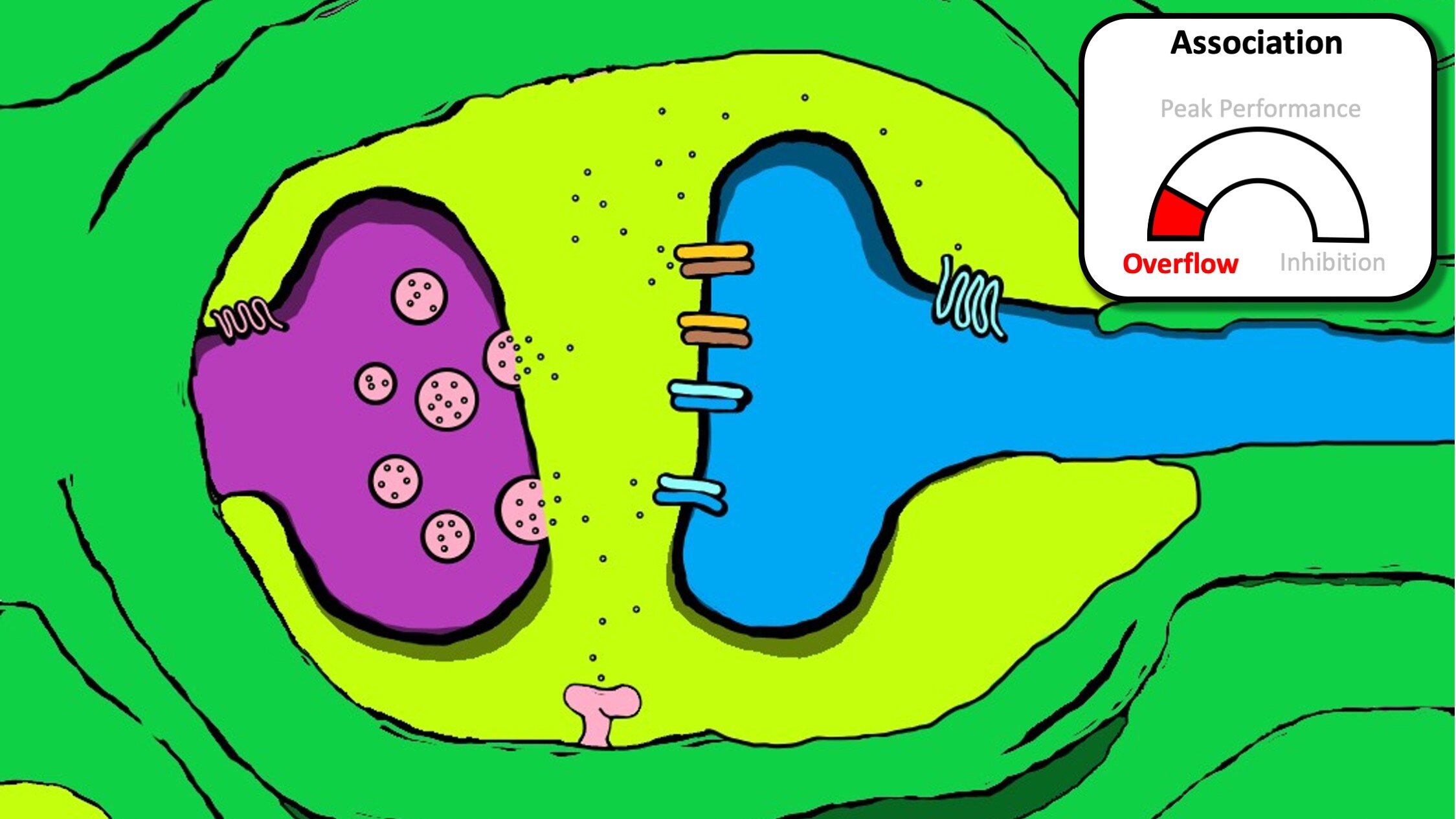
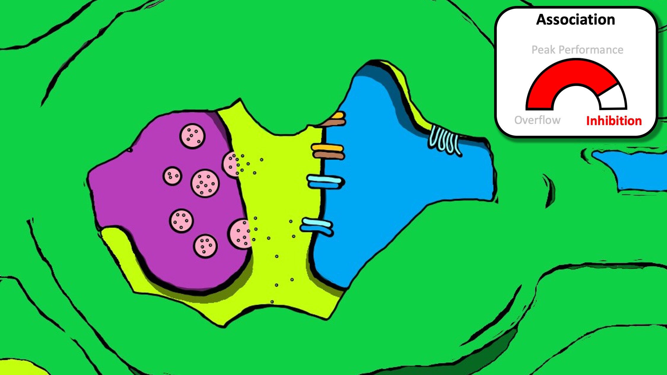
Here I switched from illustrator to MSpaint. I applied colors like paint and created a sort of more cartoon-like appearance. This figure was aimed at conveying the idea that astrocyte physical association with synapses can serve as a means regulating glutamate signals through alteration of the overall space that neurotransmission occurs in. This particular example was the model of how we anticipated astrocyte interaction with synapses in the nucleus accumbens impacted neural communication during relapse. This has not been published and is more of a fun cartoon for talks / seminars. Its a bit over simplified, of course astrocytes could coordinate more specific activation of presynaptic autoreceptors or metabotropic receptors (not shown). However, I like the organic feel the drawings have.
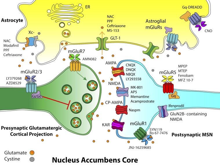
For this one I constructed a schematic of the tripartite synapse with all kinds of receptors. This figure aims to represent the corticostriatal synapse. In addition to the receptors present, Ive listed drugs that target these receptors that are capable of preventing relapse. The original publication can be found here.

Here we have a graphical representation of neuronal and astroglial mechanisms for expression of extracellular proteases linked to addiction and relapse. This was done in collaboration with another great friend and colleague of mine Alex Smith. This one ended up in a review article on ECM signaling. The original publication can be viewed here.

This is another view of a synapse and adjacent astrocyte. Here, the extracellular matrix and corresponding signaling relevant to relapse is described. The numbered aspects of the signaling cascade depict aspects of relapse biology that lead to the transient synaptic plasticity that underlies cued cocaine seeking. The original article can be found here.

Here is an early collaboration figure I did my great friend and colleague Jasper Heinsbroek while in the Kalivas lab. My greatest regret for this one is that the color blends depicting the extent of innervation of the accumbens core vs the shell was missed / unclear. This was part of a large we review project we undertook together. The original publication can be found here.

Here is another older powerpoint cartoon I put together attempting to compile and illustrate means by which glutamate can be released by astrocytes. This one is function over form for sure. Everything is a bit boxy. This schematic was part of a larger review aimed at understanding how drugs negatively impact astrocyte function and how this is linked to addiction. The original article can be found here.

This is a super early figure I attempted as a graduate student. Looking at this one I feel half nostalgic and half embarrassed of my early attempts at graphic design. Its pretty basic and details stoichiometric aspects of subunit combinations in nicotinic acetylcholine receptors. This was done with powerpoint all the way back during my graduate studies with Paul Gardner at UMass Medical. I remember being really excited about the “plasma membrane” representation. It seems pretty bland by todays standards. The full publication can be found here.